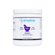
Simplesa AAKG+ Core Powder Nutrients Support Parkinson’s Disease
15th Jun 2015
The ingredients in Simplesa AAKG+ Core Powder have been shown to exhibit benefits in several neurodegenerative diseases, including Parkinson’s disease. This paper details how the combination of Arginine Alpha-Ketoglutarate, GABA, Ubiquinol and Niacin found in Simplesa AAKG+ Core Powder, along with Liposomal Glutathione, may enhance mitochondrial function and be effective stand alone and co-therapies in the management of Parkinson’s disease.
Click here for more information on Simplesa AAKG+ Core Powder.
The ingredients in Simplesa AAKG+ Core Powder have been shown to benefit several neurodegenerative diseases including Parkinson’s disease. This paper details how the combination of Arginine Alpha-Ketoglutarate, GABA, Ubiquinol and Niacin found in Simplesa AAKG+ Core Powder, along with Glutathione, may enhance mitochondrial function and be an effective stand alone and co-therapy in the management of Parkinson’s disease.
Parkinson’s Disease: An Overview and Additional Insights
Parkinson's disease (PD) is the second most common neurodegenerative disorder after Alzheimer's disease, affecting approximately 1% of the population older than 50 years. There is a worldwide increase in disease prevalence due to the increasing age of human populations (1). PD is an idiopathic disease of the nervous system characterized by both motor and non-motor system manifestations. It is a chronic progressive neurodegenerative disorder that occurs mostly in older persons but can also appear in much younger patients. Other neurodegenerative disorders can mimic idiopathic PD, including Dementia with Lewy Bodies (DLB), Corticobasal Degeneration (CBD), Multiple System Atrophy (MSA) and Progressive Supranuclear Palsy (PSP). However, the major focus of this review will be idiopathic (unknown cause) PD and not these other Parkinsonian-like syndromes (2).
Symptom picture
Parkinson’s disease causes four main symptoms including slowed movement (bradykinesia), postural instability, muscle rigidity, and a resting tremor (3). Other secondary motor symptoms that are often present include shuffling gait, freezing, and dystonia (repetitive movements) (4). Non-motor symptoms include anxiety, depression, sweating, memory impairment, sexual dysfunction, sleep disturbances, impulse control, bladder issues, and constipation (5).
Pathophysiology
Parkinson’s disease is primarily associated with the gradual loss of cells in the substantia nigra of the brain. This area is responsible for the production of dopamine which is a chemical messenger (neurotransmitter) that transmits signals between two regions of the brain to coordinate activity. Dopamine allows the substantia nigra and the corpus striatum to communicate to regulate muscle activity.
If there is deficiency of dopamine in the striatum the nerve cells in this region “fire” out of control. This leaves the individual unable to direct or control movements. This leads to the initial symptoms of Parkinson’s disease. As the disease progresses, other areas of the brain and nervous system degenerate as well, causing a more profound movement disorder. A definitive neuropathological diagnosis of Parkinson's disease requires loss of dopaminergic neurons in the substantia nigra and related brain stem nuclei and the presence of Lewy bodies in remaining nerve cells (6). Lewy bodies are abnormal aggregates of alpha synuclein protein that develop inside nerve cells in Parkinson's disease (PD), Lewy body dementia, and some other disorders.
Genetic mutations, neuro inflammation, oxidative stress, and the aging process have all been implicated in the pathogenesis of PD (7, 8, and 9). Probable causes also include mitochondrial dysfunction, misfolded protein damage, alteration of cellular clearance systems, abnormal calcium handling and altered inflammatory response, all represent key targets for neuroprotection (10). Reduced levels of cofactors for cellular mitochondrial energy production have been found in the majority of patients with PD (8).
The contribution of genetic factors to the pathogenesis o f Parkinson's disease is increasingly being recognized. A point mutation which is sufficient to cause a rare autosomal dominant form of the disorder has been recently identified in the alpha-synuclein gene on chromosome 4 in the much more common sporadic, or 'idiopathic' form of Parkinson's disease, and a defect of complex I of the mitochondrial respiratory chain was confirmed at the biochemical level. Diseases specificity of this defect has been demonstrated for the parkinsonian substantia nigra (6).
Neuro inflammation constitutes a fundamental process involved in the physiopathology of Parkinson's disease (PD). Microglial cells (immune cells of the brain) play a central role in the outcome of neuro inflammation and consequent neurodegeneration of dopaminergic neurons in the substantia nigra (7), suggesting there is an autoimmune component to PD. As autoimmunity is often the result of poor detoxification, it is highly probable that enhancement of mitochondrial function and reduction of oxidative stress would greatly improve outcomes in patients with Parkinson’s disease.
Current Therapies and Future Considerations
There are three main categories of pharmaceutical drugs that help manage the symptoms of PD. L-dopa is the cornerstone of Parkinson's drug therapy. It helps replenish the brain's supply of dopamine, reducing the tremors and other motor symptoms of Parkinson's. It typically helps most with bradykinesia and rigidity, but not balance problems. L-dopa usually is combined with the drug carbidopa to diminish nausea, vomiting, and other side effects. Although there is often noticeable improvement after starting L-dopa, people typically need to gradually increase the dose for maximum benefit (11).
Dopamine agonistsmimic dopamine in the brain and can be given alone or with L-dopa. They include apomorphine, pramipexole, ropinirole, and rotigotine. Selegiline (also called deprenyl) and rasagiline are MAO-B inhibitors which cause dopamine to accumulate in surviving nerve cells and reduce the symptoms of Parkinson's. Entacapone and tolcapone are COMT inhibitors which prolong L-dopa's effects by preventing the breakdown of dopamine. The last group of drugs are anticholinergics which are not PD specific and are often helpful in the treatment of tremors. All of these therapies will not treat the entirety of symptoms in Parkinson’s and often decrease in efficacy as the length of use increases (11, 12).
Deep brain stimulation for PD entered widespread clinical use in the late 1990s and acceptance of this therapy has increased over the past 15 years. The attractiveness of deep brain stimulation is related in part to the fact that stimulation is adjustable and reversible. However, recent reports highlighting unexpected behavioral effects of stimulation suggest that deep brain stimulation, while improving motor function, may have other less desirable consequences. The popularity of deep brain stimulation belies the fact that its utility relative to medical therapy has been studied only in uncontrolled studies, with 1 recent exception. The best candidates for this are those with moderate to severe PD with motor complications responsive to levodopa and no significant cognitive impairment (12).
Progressive neuronal cell loss in a small subset of brainstem and mesencephalic nuclei and widespread aggregation of the α-synuclein protein in the form of Lewy bodies and Lewy neurites are neuropathological hallmarks of Parkinson's disease. Most cases occur sporadically, but mutations in several genes, including SNCA, which encodes α-synuclein, are associated with disease development. The discovery and development of therapeutic strategies to block cell death in Parkinson's disease has been limited by a lack of understanding of the mechanisms driving neurodegeneration. However, increasing evidence of multiple pivotal roles of α-synuclein in the pathogenesis of Parkinson's disease has led researchers to consider the therapeutic potential of several strategies aimed at reduction of α-synuclein toxicity (13). Phenserine or Posiphen reached clinical assessment for PD as an anticholinesterase and blocker of amyloid precursor protein translation (anti-amyloid). This well-tolerated compound also reduced neural alpha-synuclein expression over a similar dose range. Researchers will pursue posiphen (stereoisomer of phenserine) for its capacity to lower alpha-synuclein expression, first in cell culture, then progressing to animal models (14). Another compound that reduces alpha-synuclein cytotoxicity (or neuronal cell death) is curcumin. This is just one natural substance under investigation or already in use for the treatment or adjunct therapy for Parkinson’s disease (15).
Alternative and Adjuvant Therapies
Due to the role oxidative stress plays in the pathogenesis of PD, several vitamins and neuromodulators that enhance mitochondrial function have gained ground as promising and effective stand alone and co-therapies in the management of Parkinson’s disease. Replacement of cellular energy cycle cofactors that are often found to be deficient in PD sufferers has been shown to be highly efficacious in restoring positive response to dopamine therapies. Recent studies have shown that midbrain dopaminergic neurons release other neurotransmitters other than just dopamine (16).
GABA
CSF GABA levels are decreased in PD patients treated with levodopa who are unresponsive as well as in untreated PD patients (17). Those who no longer respond to dopamine therapies respond once more when the other neurotransmitters, especially GABA, which is found to be produced by dopaminergic neurons, are supplemented as well (16). Low levels of GABA contribute to muscle spasticity and stiffness and replacement of GABA can not only help reduce this symptom, but many others associated with PD due to its synergistic relationship to dopamine.
Arginine Alpha-ketoglutarate
Alpha-ketoglutarate dehydrogenase is also a major participant in mitochondrial function and neurodegenerative disease. There are reduced levels of alphaketoglutarate dehydrogenase in the brains of people with Parkinson’s disease. Alpha-ketoglutarate is an important substrate for the TCA/Krebs cyclewhich drives cellular energy processes. A lack of AKG stops the Krebs cycle and the affected cell dies. This applies to neuronal cells such as dopaminergic cells as well. 1-methyl-4-phenyl-1,2,3,6-tetrahydropyridine (MPTP) is a neurotoxin associated with drug abuse and causes permanent symptoms of Parkinson's disease(PD) by destroying dopaminergic neurons in the substantia nigra of the brain. Treatment with Alpha-ketoglutarate were found to resolve the loss of motor coordination, oxidative stress, diminished complex I activity and tyrosine hydroxylase-positive neurons in midbrain (18).
Ubiquinol/CoQ10
CoQ10 is another substrate that is found to be deficient in patients with PD and is also able to attenuate the MPTP-induced loss of striatal dopaminergic neurons. Variants of CoQ10 play a role in enzyme complexes I-V that function in oxidative phosphorylation. A defect in mitochondrial oxidative phosphorylation, in terms of a reduction in the activity of NADH CoQ reductase (complex I) has been reported in the striatum of patients with Parkinson's disease. The reduction in the activity of complex I is found in the substantia nigra, but not in other areas of the brain, such as globus pallidus or cerebral cortex. Therefore, the specificity of mitochondrial impairment may play a role in the degeneration of nigrostriatal dopaminergic neurons. This view is supported by the fact that MPTP generating 1-methyl-4-phenylpyridine (MPP(+)) destroys dopaminergic neurons in the substantia nigra (1). Oral CoQ10 was found to protect the nigrostriatal dopaminergic system in one-year-old mice treated with MPTP, a toxin injurious to the nigrostriatal dopaminergic system. Oral CoQ10 was well absorbed in parkinsonian patients and caused a trend toward increased complex I activity. These data suggest that CoQ10 may play a role in cellular dysfunction found in PD and may be a potential protective agent for parkinsonian patients (19).
Niacin/Nicotinamide
Nicotinamide, the principal form of niacin (vitamin B3), has been proposed to be neuroprotective in Parkinson's disease. Niacin-derived NAD(P)H as antioxidants and enzyme cofactors can inhibit oxidative damage and improve mitochondrial function and thus protect neurodegeneration and improve motor function according to a study done on an alpha-synuclein transgenic Drosophila Parkinson's model. Mitochondrial function was tested by measuring the activity of mitochondrial complex I and alpha-ketoglutarate dehydrogenase, and oxidative damage was tested by measuring reactive oxygen species, DNA damage, and protein oxidation levels. Nicotinamide at a relatively higher concentration significantly protected human neuroblastoma cells from an MPP(+)-induced decrease in cell viability, complex I and alpha-ketoglutarate dehydrogenase activity, and an increase in oxidant generation, DNA damage, and protein oxidation. . These results suggest that nutritional supplementation of nicotinamide at high doses decreases oxidative stress and improves mitochondrial and motor function in cellular and/or Drosophila models and may be an effective strategy for preventing and ameliorating Parkinson's disease (20).
Glutathione
Neuronal loss in the substantia nigra is heavily correlated with oxidative stress on a cellular level. Reduction of oxidative stress has been shown to slow and in some cases halt the destruction of the substantia nigra and dopaminergic neurons. One of the most powerful antioxidants available is glutathione. Several studies have shown that glutathione is not only neuroprotective but it is abnormally low in those with PD (20, 21, 22, 23).
Click here for more information on Simplesa AAKG+ Core Powder.
References
1.Ebadi M, et al. Ubiquinone (coenzyme q10) and mitochondria in oxidative stress ofParkinson’s disease. Biol Signals Recept.2001 May-Aug; 10(3-4):224-53.
2.Beitz, Janice. Parkinson's disease: a review. Frontiers in Bioscience. S6, (65-74), January 1, 2014.
3.Weiner, William J. Parkinson's Disease: A Complete Guide for Patients and Families.The Johns Hopkins University Press: October 8, 2013; (4-5).
4.Jancovic J. Parkinson’s disease: clinical features and diagnosis. J Neurol Neurosurg Psychiatry2008;79:368-376doi:10.1136/jnnp.2007.131045
5.Calleo JS, et al. A Pilot Study of a Cognitive-Behavioral Treatment for Anxiety and Depression in Patients withParkinson Disease. J Geriatr Psychiatry Neurol.2015 Jun 4. pii: 0891988715588831.
6.Ebadi M, et al. Ubiquinone (coenzyme q10) and mitochondria in oxidative stress ofParkinson’s disease. Biol Signals Recept.2001 May-Aug;10(3-4):224-53.7. H, et al. Regulation of the Neurodegenerative Process Associated toParkinson's Diseaseby CD4+ T-cells. J Neuroimmune Pharmacol.2015 May 28. [Epub ahead of print]
8.Gibson GE, Kingsbury AE, Xu H, et al. Deficits in a tricarboxylic acid cycle enzyme in brains from patients with Parkinson’s disease. Neurochemistry"International."2003; 43 (2):129D135. 129.
9.Jia H, et al. High doses of nicotinamide prevent oxidative mitochondrial dysfunction in a cellular model and improve motor deficit in a Drosophila model ofParkinson's disease. J Neurosci Res.2008 Jul;86(9):2083-90. doi: 10.1002/jnr.21650.
10.De Rosa P, et al. Candidate genes forParkinson disease: Lessons from pathogenesis. Clin Chim Acta.2015 Jun 2. pii: S0009-8981(15)00277-6. doi: 10.1016/j.cca.2015.04.042. [Epub ahead of print]
11.NIH. Parkinson's Disease: Diagnosis and Treatment. Winter2014Issue: Volume8Number4Page8-10
12.Weaver F, Follett K, Hur K, Ippolito D, Stern M. Deep brain stimulation in Parkinson disease: a metaanalysis of patient outcomes. J Neurosurg. 2005;103(6):956-967
13.Dehay B, et al. Targeting α-synuclein for treatment ofParkinson'sdisease: mechanistic and therapeutic considerations. Lancet Neurol.2015 Jun 3. pii: S1474-4422(15)00006-X. doi: 10.1016/S1474-4422(15)00006-X. [Epub ahead of print]
14.https://www.michaeljfox.org/foundation/grant-detail.php?grant_id=402
15.Wang MS, et al. Curcumin reduces alpha-synuclein induced cytotoxicity inParkinson'sdiseasecell model. BMC Neurosci.2010 Apr 30;11:57. doi: 10.1186/1471-2202-11-57.
16.Tritsch NX, Ding JB, Sabatini BL. DOPAMINERGIC NEURONS INHIBIT STRIATAL OUTPUT THROUGH NON-CANONICAL RELEASE OF GABA. Nature. Oct 11, 2012;490(7419):262-
17.Félix J. Jiménez-Jiménez, et al. Cerebrospinal fluid biochemical studies in patients with Parkinson's disease: toward a potential search for biomarkers for this disease. Front Cell Neurosci. 2014; 8: 369.
18.Satpute R, et al. Neuroprotective effects of α-ketoglutarate and ethyl pyruvate against motor dysfunction and oxidative changes caused by repeated 1-methyl-4-phenyl-1,2,3,6 tetrahydropyridine exposure in mice. Hum Exp Toxicol.2013 Jul;32(7):747-58. doi: 10.1177/0960327112468172.
19.Shults CW1,Haas RH,Beal MF. A possible role of coenzyme Q10 in the etiology and treatment ofParkinson's disease. Biofactors.1999;9(2-4):267-72.
20.Jia H, et al. High doses of nicotinamide prevent oxidative mitochondrial dysfunction in a cellular model and improve motor deficit in a Drosophila model ofParkinson's disease. J Neurosci Res.2008 Jul;86(9):2083-90. doi: 10.1002/jnr.21650.
21.Gu F., Chauhan V., Chauhan A. Glutathione redox imbalance in brain disorders.Current Opinion in Clinical Nutrition and Metabolic Care.2015;18:89–95
22.De Blundel D., Schallier A., Loyens E., Fernando R., Miyashita H., Van Liefferinge J., Vermoessen K., Bannai S., Sato H., Michotte Y., Smolders I., Massie A. Loss of system xc- formula does not induce oxidative stress but decreases extracellular glutamate in hippocampus and influences spatial working memory and limbic seizure susceptibility.Journal of Neuroscience.2011;31:3592–5803
23.Dringen R., Pfeiffer B., Hamprecht B. Synthesis of the antioxidant glutathione in neurons: supply by astrocytes of CysGly as precursor for neuronal glutathione.Journal of Neuroscience.1999;19(2):562–569


